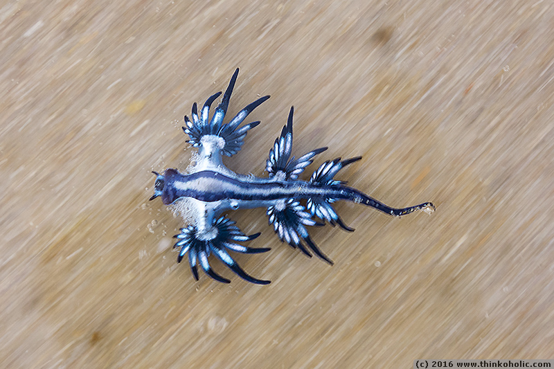
blue dragon: a beautiful bad-ass nudibranch
on a recent beach stroll on the northern new south wales coast, i came across small weird-looking blue-and-white creatures with lots of appendages. having washed ashore, they looked more like […]
a blog by markus nolf
on a recent beach stroll on the northern new south wales coast, i came across small weird-looking blue-and-white creatures with lots of appendages. having washed ashore, they looked more like […]
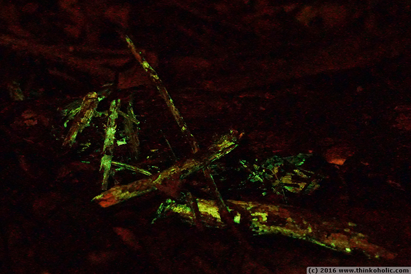
i’ve always been fascinated by bioluminescent phenomena. in a relatively wide range of organisms, evolution at some point produced species that can produce light, from marine plankton in warm seas, […]
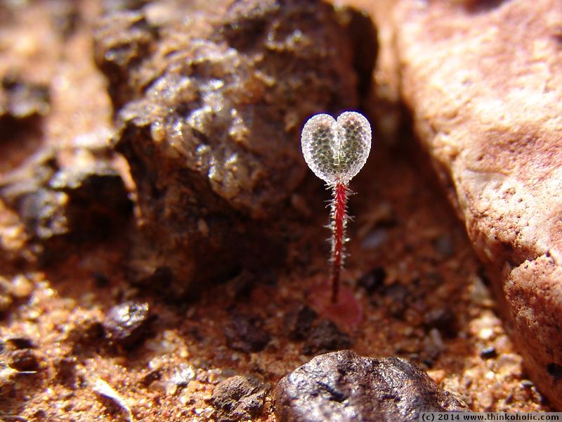
the tiny plant below is a stone plant seedling (aizoaceae), growing in the harsh conditions of the “knersvlakte” quartz gravel landscape in north-eastern south africa. the seedling has only formed […]
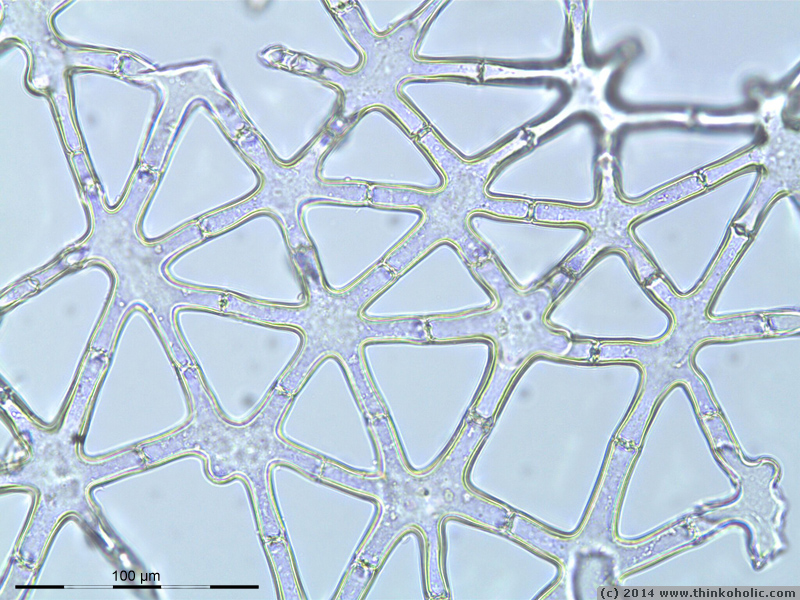
stellate parenchyma is a form of aeration tissue (aerenchyma) in plants, which helps with internal air circulation in plants. the tissue is typical of aquatic and wetland plants, and consists […]
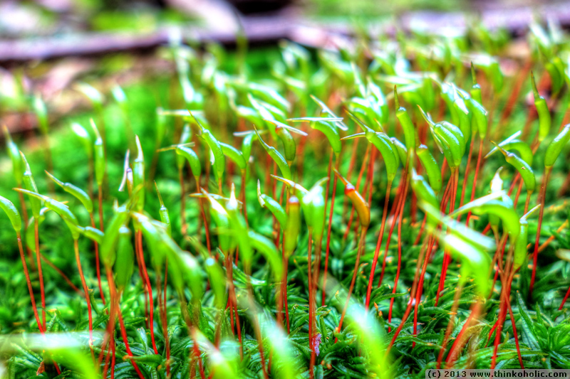
the common smoothcap moss (atrichum undulatum) features transversely undulate leaves, but its most prominent feature are the long, distinctly beaked capsules that are borne on 3 cm long, reddish setae. […]
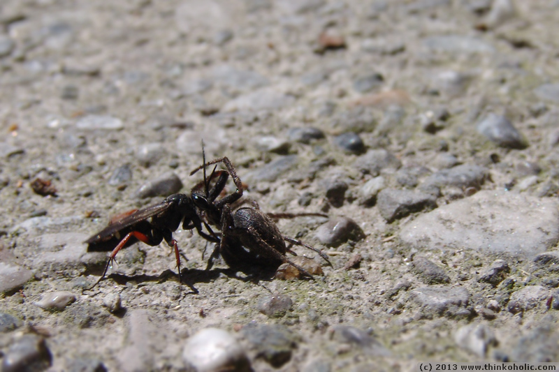
spider wasps (pompilidae) are – in a nutshell – really badass insects. to provide food for their larvae, these wasps go hunting for spiders that are usually bigger than themselves. […]