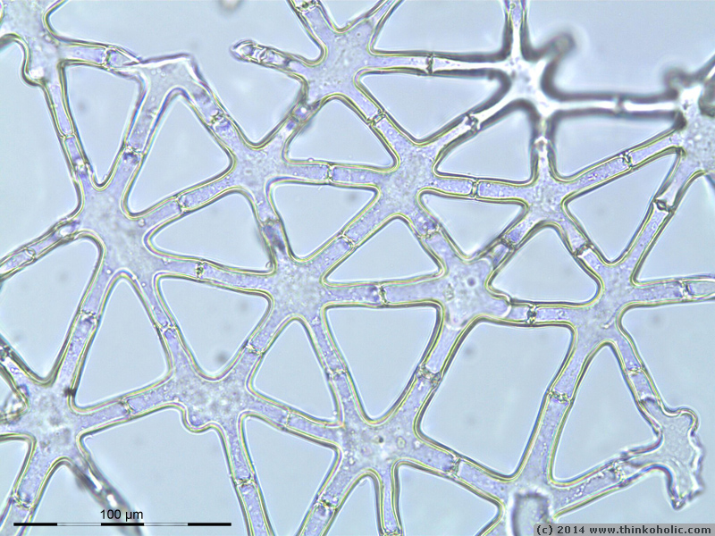
stellate parenchyma [photo and animation]
stellate parenchyma is a form of aeration tissue (aerenchyma) in plants, which helps with internal air circulation in plants. the tissue is typical of aquatic and wetland plants, and consists […]
a blog by markus nolf
stellate parenchyma is a form of aeration tissue (aerenchyma) in plants, which helps with internal air circulation in plants. the tissue is typical of aquatic and wetland plants, and consists […]
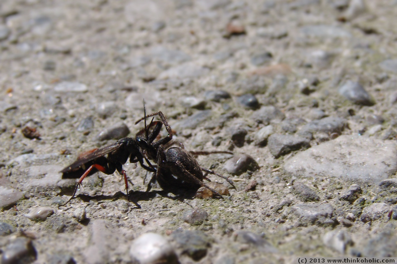
spider wasps (pompilidae) are – in a nutshell – really badass insects. to provide food for their larvae, these wasps go hunting for spiders that are usually bigger than themselves. […]
this little animation shows the development of an anatomical drawing that i did during a zoology class a few years ago. the reconstruction is part of a university project for […]
this animation was done as a request. a friend needed such a file for her presentation, but couldn’t find one on the internets… animation: a venus flytrap (dionaea muscipula) closes […]
nature often keeps me busy concentrating to not just stare at something with an open mouth. in a recent class, we looked at developmental stages of plant embryos, from the […]
browsing through the photos from vienna, i found two very similar pictures of the church of st. charles in vienna – one taken in daylight, the other one 2.5 hours […]